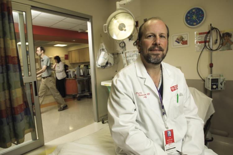
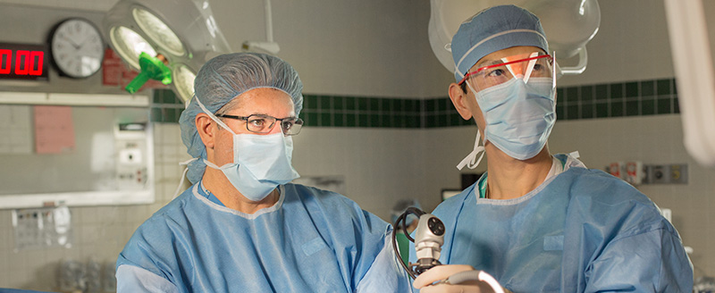
Chicago Institute of Neurosurgery and Neuroresearch (CINN) Brain Tumor Program
CINN treats a wide range of neuro-related pain disorders, from common back and neck pain, to facial pain, to various types of headaches. Non-surgical solutions for these conditions are managed through the physiatry and neurology teams at CINN. These practitioners have specialized expertise in utilizing an array of therapies, medications and other non-surgical means to control tangoporno pain. Unfortunately, in some circumstances these non-invasive or minimally invasive therapies are not effective and surgical options, including Gamma Knife stereotactic radiosurgery, should be explored. At that point patients are referred to a highly trained, subspecialized, CINN neurosurgeon who might be able to provide a surgical solution.
The following section of the CINN website provides an overview of back and neck pain, facial pain and headaches, along with the various treatment options available through CINN. If you have further questions regarding our services, or would like to schedule an appointment with a CINN physician, please call 1-800-446-1234.
Neck pain
Neck pain accounts for almost 1% of all visits to a primary care physician in the United States and may result from abnormalities in the soft tissues – the muscles, ligaments, and nerves – as well as in bones and joints of the spine.
Low Back pain
Ninety percent of lower back pain (LBP) cases are benign and typically subside within six weeks regardless of treatment method. The diagnosis and treatment of the remaining 10 percent are more challenging, however, and demand strict adherence—both from the physician and the patient—to the latest clinical rubias19 guidelines and recommendations. Based on current literature, a multidisciplinary approach combining drug treatment with physical and patient education has proven to be the most successful treatment of back pain.
Welcome to the website for the Chicago Institute of Neurosurgery and Neuroresearch (CINN) Brain Tumor Program. Learning that you or a loved one has a brain tumor is a frightening event. The many questions and concerns about the diagnosis and possible treatments can be overwhelming. This website was developed to help you, your family and friends learn about and better understand brain tumors. It will also help you understand how CINN’s dedicated team approaches treatment.
Brain tumors are a complex mass of abnormal cells in the brain of youporn. A difficult disease, the symptoms can be quite debilitating. The CINN Brain Tumor Program offers a coordinated, interdisciplinary approach to the care of adult and pediatric brain tumor patients. Our team of more than 200 full-time staff includes physicians, physician assistants, nurses, neuro-psychologists, physical, occupational and speech therapists, social workers and clinical researchers, all who have dedicated their careers to working with brain tumor patients. We know that you realize the importance and value of this multidisciplinary approach. The specialists at CINN provide state-of-the-art treatment and are continuously refining and developing new porno and effective forms of early diagnosis and therapy. Along with improving technology and the gradual unfolding of scientific understanding of the basic biology of brain tumors, patients and families can look to the future with a considerable amount of hope.
If you would like more information or would like to see a CINN physician please call 1-800-411-CINN.
Sincerely,
The CINN Brain Tumor Program Team